Skin Growth Removal
Skin tumors are quite common and most often not dangerous. The exception is melanoma, which bears mortal danger and makes us stay alert.
Cancer vigilance: diagnosis first, removal second
We never remove a growth "by eye". Before the procedure, the doctor performs a consultation with dermatoscopy, examining the lesion under high magnification and assessing its structure, borders and colour. Digital dermatoscopy lets us photograph the lesion and compare it at follow-up visits.
Based on the results, the doctor decides whether removal is needed and which method to use. Some lesions are safer to monitor than to remove. After removal, the lesion is sent for histology, which confirms that it is benign and gives you complete peace of mind. If there is any suspicion of malignancy, we refer the patient to an oncologist and do not perform cosmetic removal.
When to see a dermatologist
— a mole starts growing, especially one you have had for years;
— its shape becomes asymmetrical or its borders irregular or blurred;
— its colour changes or becomes uneven;
— it grows larger than 5–6 mm;
— it itches, bleeds, crusts or ulcerates;
— a new, fast-growing lesion appears;
— one mole looks noticeably different from the others.
What we remove
Benign skin growths: papillomas; acrochordons (skin tags); fibromas and dermatofibromas; moles (nevi), including intradermal and epidermal nevi; seborrheic keratoses; angiomas and cherry angiomas; pyogenic granulomas; syringomas; xanthelasma; cutaneous horns; fibrous papules; sebaceous hyperplasia; nevus sebaceous.
Subcutaneous growths and cysts: lipomas; epidermoid cysts; trichilemmal (pilar) cysts; milia; dermoid cysts; pilomatricomas and other superficial benign growths of the subcutaneous tissue.
Viral growths: common, flat, plantar and filiform warts; molluscum contagiosum; genital warts (condylomas).
Other growths: injured benign lesions; lesions in difficult locations — eyelids, nasal ala, vermilion border of the lips, ears, scalp; other skin and subcutaneous growths once a diagnosis has been made.
Removal methods
We use three removal methods. The doctor chooses one individually, depending on the type, size, depth and location of the lesion.
CO2 laser (Candela CO2RE). Precise removal with controlled depth; the laser seals small vessels at the same time, so the procedure is practically bloodless. Suitable for papillomas, keratoses, warts, fibromas, syringomas, xanthelasma and sebaceous hyperplasia.
Electrocoagulation. Removal with high-frequency current that coagulates vessels at the same time. Used for small superficial lesions such as small papillomas, skin tags and angiomas.
Surgical excision. The lesion is removed completely, together with its capsule, and the wound is closed with a fine cosmetic suture. This method is chosen for lipomas, epidermoid and other cysts, and large nevi.
Whenever a lesion is sent for histology, it is removed completely within healthy tissue margins — with the CO2 laser or surgically — so that the laboratory receives a complete tissue sample.
Pain-free removal
All procedures are performed under full anaesthesia. The doctor chooses the type depending on the size, number and location of the lesions:
— topical anaesthesia — a numbing cream applied to the skin in advance;
— injection anaesthesia — local anaesthetic injected for larger and deeper lesions;
— cryoanaesthesia — cooling of the skin, convenient when removing multiple lesions in one visit.
How the procedure works
1. Doctor's consultation and dermatoscopy of the lesion, choice of removal method.
2. Anaesthesia — topical, injection or cryoanaesthesia.
3. Removal of the lesion with the CO2 laser, electrocoagulation or surgical excision — from a few minutes to half an hour, depending on size and number of lesions.
4. Antiseptic treatment of the wound; a suture and dressing if needed.
5. The removed lesion is sent to a laboratory for histology.
6. Personal aftercare advice and a follow-up visit.
Healing and results
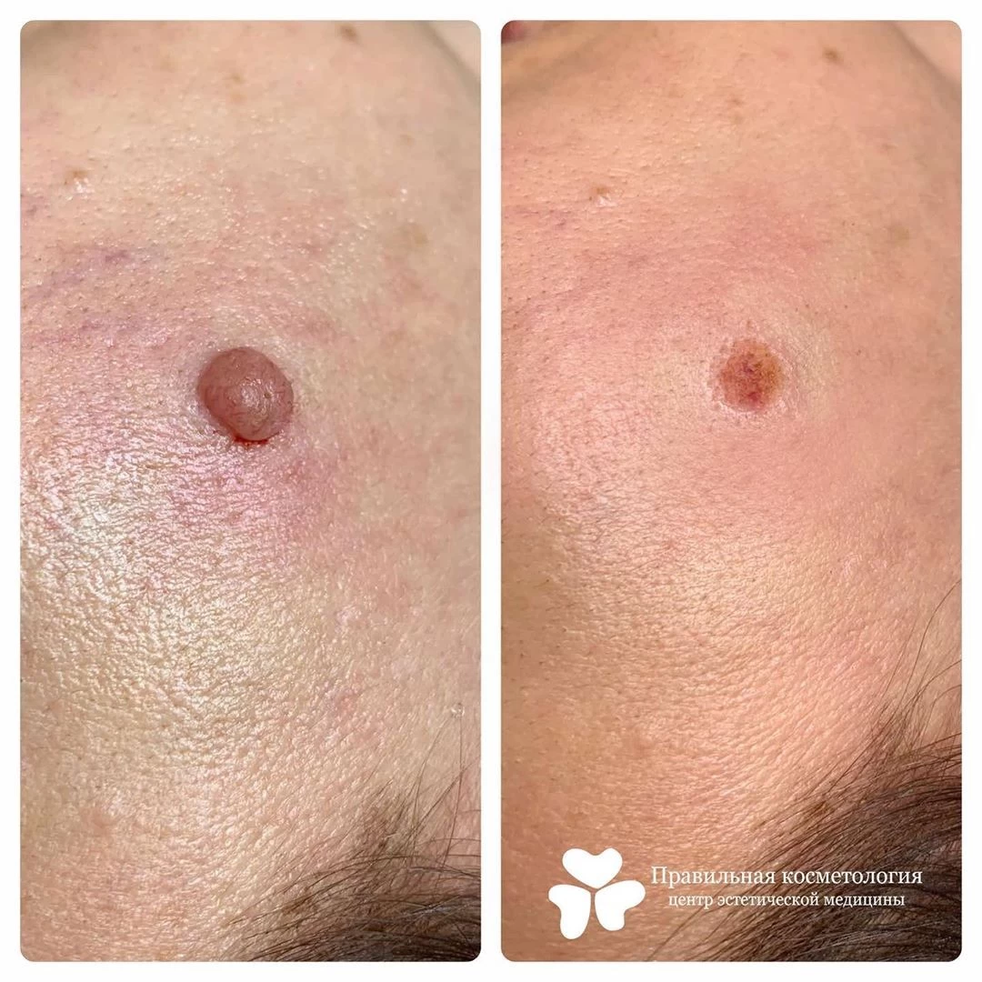
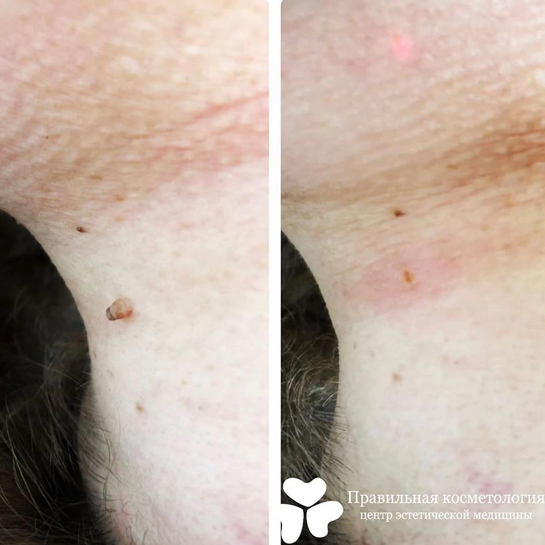
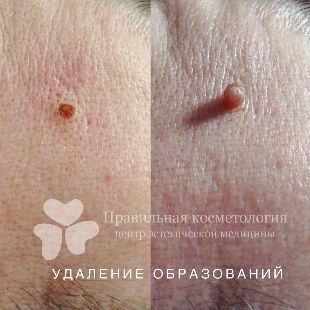
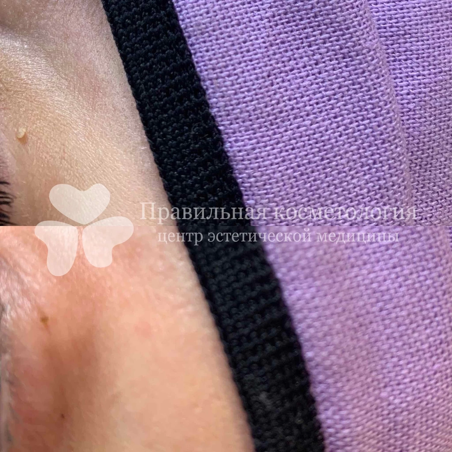
After CO2 laser removal or electrocoagulation, a thin crust forms and falls off on its own in about 5–10 days (usually faster on the face, longer on the body). After surgical excision, a cosmetic suture is placed, and the doctor will tell you when it should be removed. Do not pick or soak the crust. Until fully healed, avoid pools, saunas and make-up on the area, then protect the skin with sunscreen for several weeks.
Small growths, especially pedunculated ones, are often removed with practically no trace. Removing large or deep growths leaves a small, neat scar — the doctor will explain the expected result in advance. If a mark still bothers you, it can be corrected through our scar treatment service.
Contraindications
Acute inflammation or infection in the treatment area, active herpes, fever, decompensated chronic conditions, bleeding disorders. Elective cosmetic removal is usually postponed during pregnancy. The final decision is made by the doctor at the consultation.
Why Pravylna Kosmetolohiya
— over 20 years in Kharkiv;
— removals are performed by dermatologists with strict cancer vigilance;
— mandatory dermatoscopy before every removal;
— histology of removed lesions;
— full anaesthesia: numbing cream, local injection or cryoanaesthesia;
— three removal methods: CO2 laser, electrocoagulation and surgical excision, so we can choose the best option for each lesion;
— a gentle approach to the face and exposed areas, aiming for minimal marks.
Book a consultation with dermatoscopy — the doctor will assess the growth and suggest a safe way to remove it.
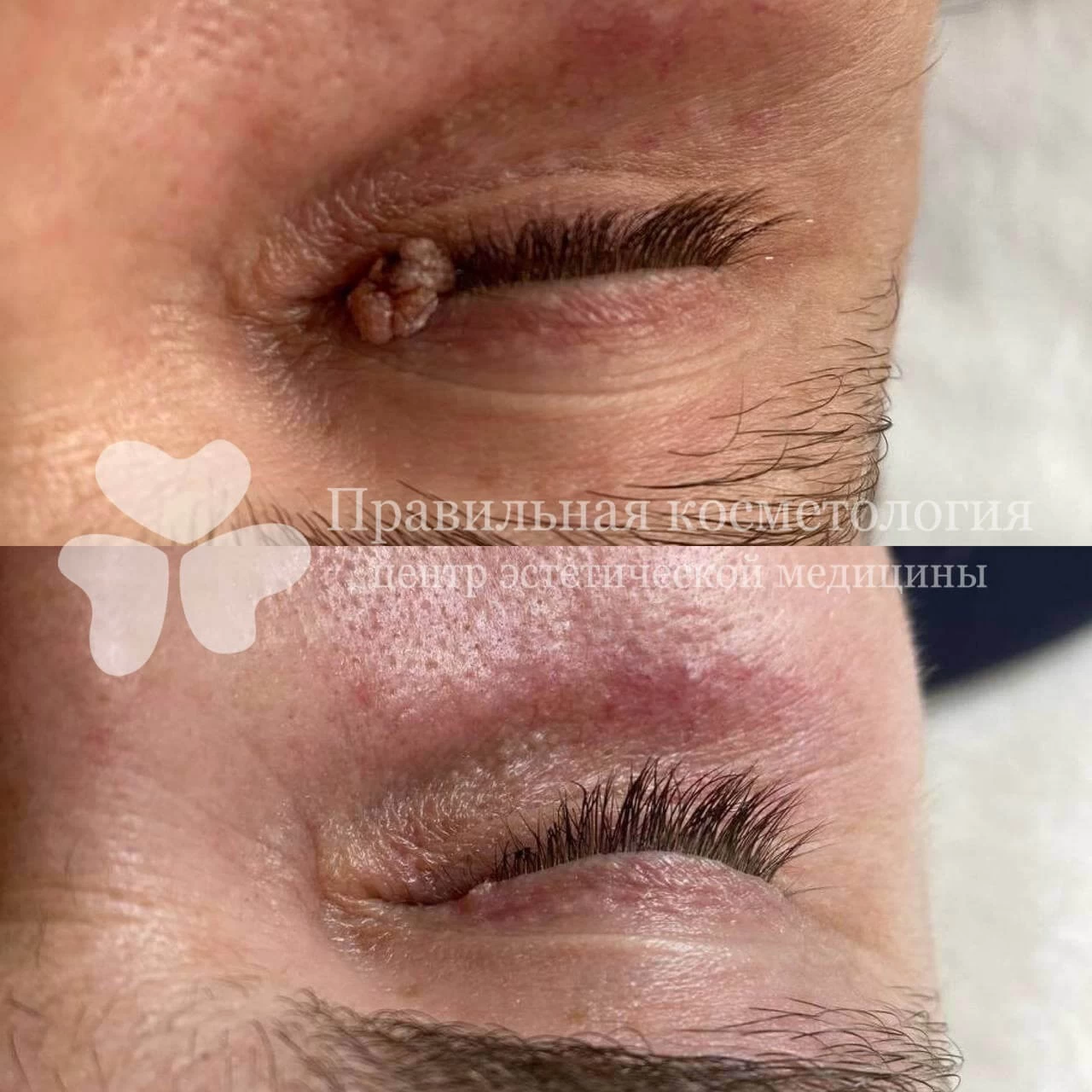
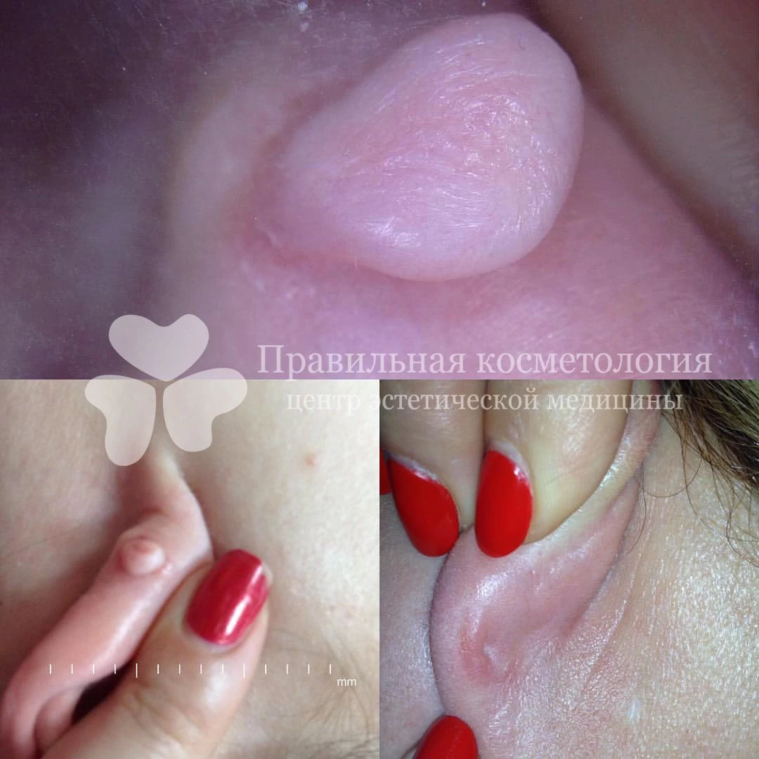
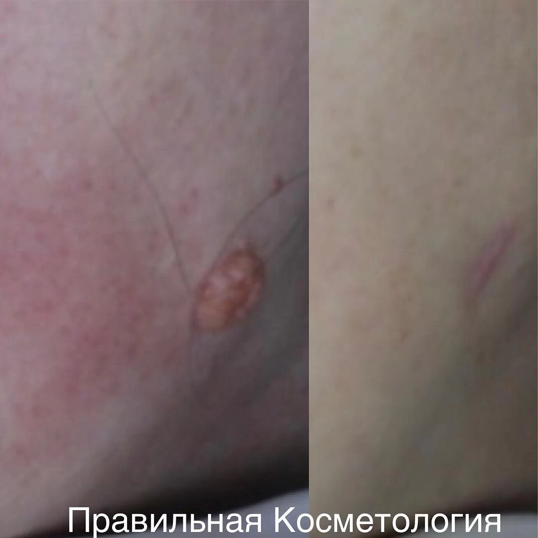
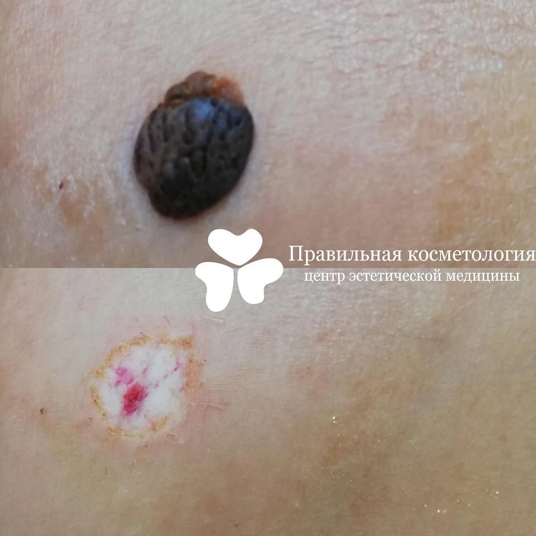
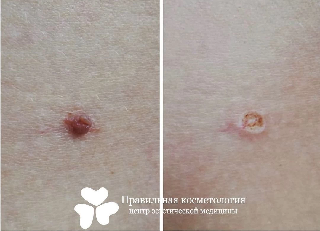
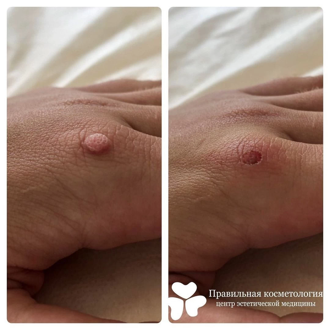
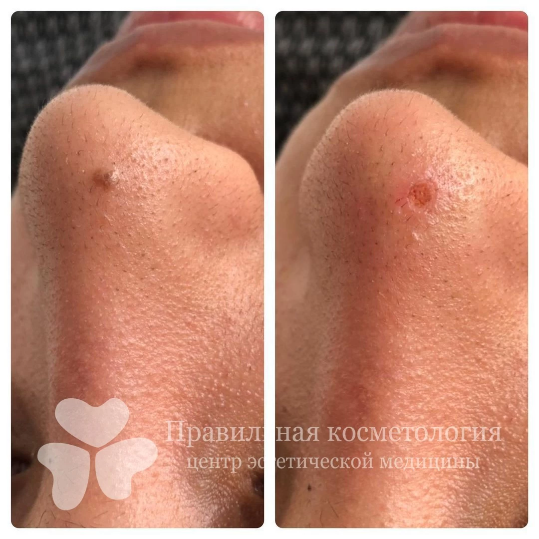
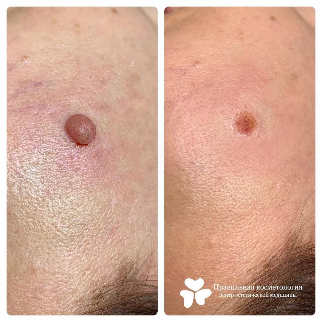
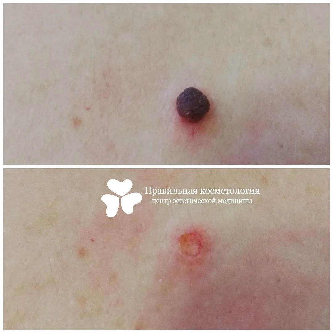
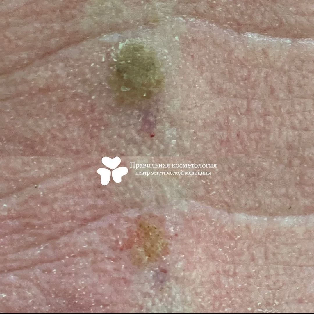
Examples of work
Video of procedures